The McKnight Endowment Fund for Neuroscience has selected four projects to receive the 2024 Neurobiology of Brain Disorders Awards. The awards will total $1.2 million for research on the biology of brain diseases, with each project receiving $100,000 per year in each of the next three years for a total of $300,000 funded per project.
The Neurobiology of Brain Disorders (NBD) Awards support innovative research by U.S. scientists who are studying neurological and psychiatric diseases. The awards encourage collaboration between basic and clinical neuroscience to translate laboratory discoveries about the brain and nervous system into diagnoses and therapies to improve human health.
An additional area of interest is the contribution of the environment to brain disorders. Early-life environmental stress is a powerful disposing factor for later neurological and psychiatric disorders. Studies show communities of color are at higher risk for these stressors, which range from environmental (e.g. climate, nutrition, exposure to chemicals, pollution) to social (e.g. family, education, housing, poverty). From a clinical perspective, understanding how environmental factors contribute to brain disease is essential for developing effective therapies.
“From expanding our understanding of how brain diseases develop to exploring novel new therapies for brain disorders, the researchers chosen for this year’s award are breaking important ground in neurological research on neurological diseases,” said Ming Guo, M.D., Ph.D., chair of the awards committee, Laurie & Steven C. Gordon Chair of Neurosciences, and Professor in Neurology & Pharmacology at UCLA David Geffen School of Medicine. “They are studying the underpinnings of devastating and life-altering conditions, including malignant brain tumors, Parkinson’s Disease and Alzheimer’s Disease, advancing ideas that could lead to new insights into how the brain works and potentially identify cures for currently incurable neurological disorders in the future.”
The awards are inspired by the interests of William L. McKnight, who founded the McKnight Foundation in 1953 and wanted to support research on brain disease. His daughter, Virginia McKnight Binger, and The McKnight Foundation board established the McKnight neuroscience program in his honor in 1977.
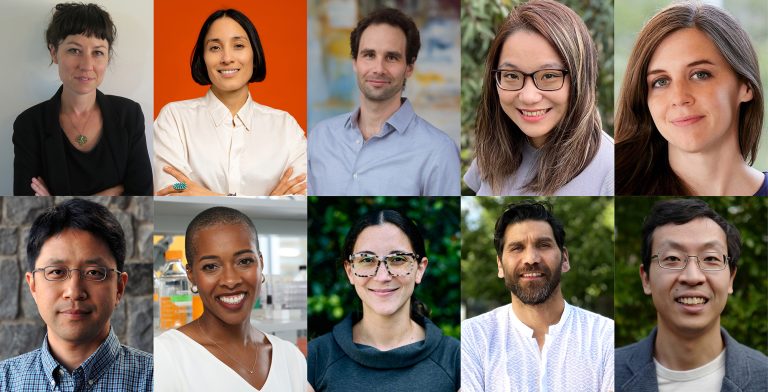
Multiple awards are given each year. This year’s four awards are:
With 134 letters of intent received this year, the awards are highly competitive. A committee of distinguished scientists reviews the letters and invites a select few researchers to submit full proposals. In addition to Dr. Guo, the committee includes Susanne Ahmari, M.D., Ph.D., University of Pittsburgh School of Medicine; Gloria Choi, Ph.D., Massachusetts Institute of Technology; André Fenton, Ph.D., New York University; Joseph G. Gleeson, M.D., University of California San Diego; Tom Lloyd, M.D., Ph.D., Baylor College of Medicine; and Michael Shadlen, M.D., Ph.D., Columbia University.
The application for Letters of Intent for the 2025 awards opens July 30, 2024.
ስለ McKnight የተፈ ሰጭ ገንዘብ ለኒውሮሳይንስ ፈንድ
የ McKnight Endowment Fund ፎርኒቫይሳይቨን በሜኒንፖሊስ, ሚኔሶታ ውስጥ በሚክክኝንት ፋውንዴሽን ብቻ የተደገፈ እና በሀገሪቱ ውስጥ ባሉ ታዋቂ የነርቭ ሳይንቲስቶች አማካይነት ይመራ ነበር. የ McKnight ተቋም ከ 1977 ዓ.ም ጀምሮ የነርቭ ሳይንስ ምርምርን አጉልቷል. ፋውንዴሽን በ 3 ዎቹ ካምፓኒዎች ቀደምት መሪዎች ከሆኑት ከዊሊየም ማክኪንሰን (1887-1978) አንዱን ዓላማ ለማከናወን ፋውንዴሽን የመዋጮ ፈንድ እ.ኤ.አ. 1986 አቋቋመ.
In addition to the Neurobiology of Brain Disorders Awards, the endowment fund also provides annual award funding through the McKnight Scholar Awards, supporting neuroscientists in the early stages of their research careers.
የአንጎል በሽታ ነርቭ በሽታ ሽልማቶች የነርቭ ሕክምና
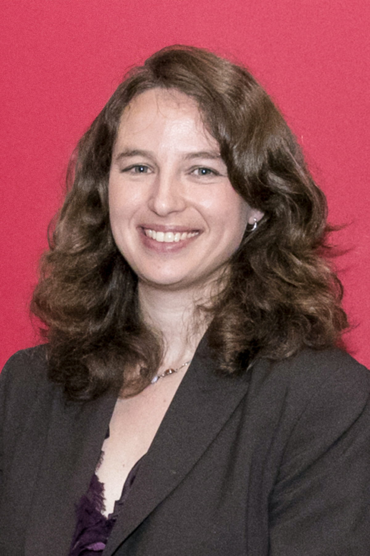
Aparna Bhaduri, Ph.D., Assistant Professor, Biological Chemistry, and co-principal investigator Kunal Patel, M.D., Neurosurgery, University of California – Los Angeles
Characterizing the Context: The Role of the Microenvironment in Shaping Human Glioblastoma:
The prognosis for people diagnosed with glioblastoma, a form of primary brain cancer, has changed very little in decades. One challenge has been that the mechanism by which glioblastoma develops and spreads is poorly understood. Mouse models can only tell researchers so much, and studies of tumors removed from the brain don’t show how it grew.
Dr. Bhaduri’s lab studies how the brain develops and how certain cell types are reactivated in the case of brain cancer, mimicking stages of brain development but coopted by the tumor. Partnering with Dr. Patel, a neurosurgeon specializing in glioblastoma surgeries, Bhaduri’s lab will use novel approaches to create systems using organoids developed from stem cell lines that closely mimic the human brain environment and then implant, grow and study tumor samples Patel collects from surgical patients. Patel has developed ways to visualize the tumors that allows him to remove some of the peripheral cells that are interfacing with surrounding brain matter, of particular interest to the research.
Bhaduri’s team will explore the lineage relationships of the glioblastoma cell types – how they change as the tumor grows, and at the roles of different cells, whether in the core, periphery or any part of the tumor – and also look at how tumor cells interact with surrounding normal cells. Understanding this link between development and glioblastoma, and how the tumor interacts with its environment, may reveal ways to disrupt it.
Aryn Gittis, Ph.D., Professor, Department of Biological Sciences, Carnegie Mellon University, Pittsburgh, PA
Investigating Circuits and Mechanisms that Support Long-Lasting Recovery of Movement in Dopamine Depleted Mice
Understanding how neural circuits control movement in humans, and how to retrain those circuits after injury or damage, is the core focus of Dr. Gittis’s lab. Her new research explores ways to tap into the brain’s plasticity to help ameliorate the effects of dopamine depletion – a key characteristic of Parkinson’s Disease– and improve movement function for longer periods of time using electrical impulses.
Deep brain stimulation, in which wires implanted in the brain deliver a constant, nonspecific electrical charge, has been approved and used to help relieve symptoms of Parkinson’s Disease for some time. However, it only addresses the symptoms, which reappear immediately when the charge is turned off. Gittis’s lab aims to find exactly what neuronal pathways are required for locomotor recovery, how electrical pulses can be “tuned” to affect just these subpopulations, and how these subpopulations can be stimulated to essentially repair themselves, offering longer-lasting relief from symptoms, even without ongoing stimulation.
Preliminary work shows promise: Working with a dopamine-depleted mouse model, Gittis and her team have identified specific subpopulations of neurons in the brain stem necessary for the relief of symptoms. Excitingly, when stimulated with a pulse of carefully tuned electricity (rather than a constant flow) the cells’ activity is changed in a way that results in hours of improved mobility with no further stimulation. Her research aims to determine whether these activity changes can be made more permanent to start healing and rewiring neural circuits.
Thanh Hoang, Ph.D., Assistant Professor, Department of Ophthalmology, Department of Cell & Developmental Biology, Michigan Neuroscience Institute, University of Michigan, Ann Arbor, MI
In vivo Reprogramming of Astrocytes into Neurons for Treating Parkinson’s Disease
Neurons of the central nervous system (CNS) are crucial for coordinating body functions, yet they are highly vulnerable to injuries. When damaged, the effects can be irreversible since neurons don’t naturally repair or replace themselves. In Parkinson’s Disease, dopaminergic neurons have lost their function, depleting dopamine in the brain. Current treatments focus on relieving symptoms such as improving motor control. Dr. Hoang is taking a different approach in his research: Finding ways to reprogram endogenous glial cells in the brain into new neurons, restoring function to the brain.
Hoang’s lab has proven the concept using retinal neurons. Using a mouse model, Hoang identified genes in retinal glial cells that act as suppressors, preventing the cells from transforming into neurons. Simultaneous loss of function to those four genes led to an almost complete conversion of those glial cells into retinal neurons. His research aims to determine if the same principle can be applied to astrocytes, the most abundant type of glial cell in the CNS, which closely resemble the retinal glia from his lab’s previous research.
In his new research, Hoang aims to reach towards a therapeutic application. He is working to perfect an in vivo process to inhibit the suppressors in the astrocytes via adeno-associated virus (AAV) vector. His research will first identify the types of neurons that result from the process – many types appear to result – and then seek to determine what factors are required to promote the development and maturation of dopaminergic neurons specifically. This work promises to advance the science of cell reprogramming, with implications for many neurological disorders in addition to Parkinson’s Disease.
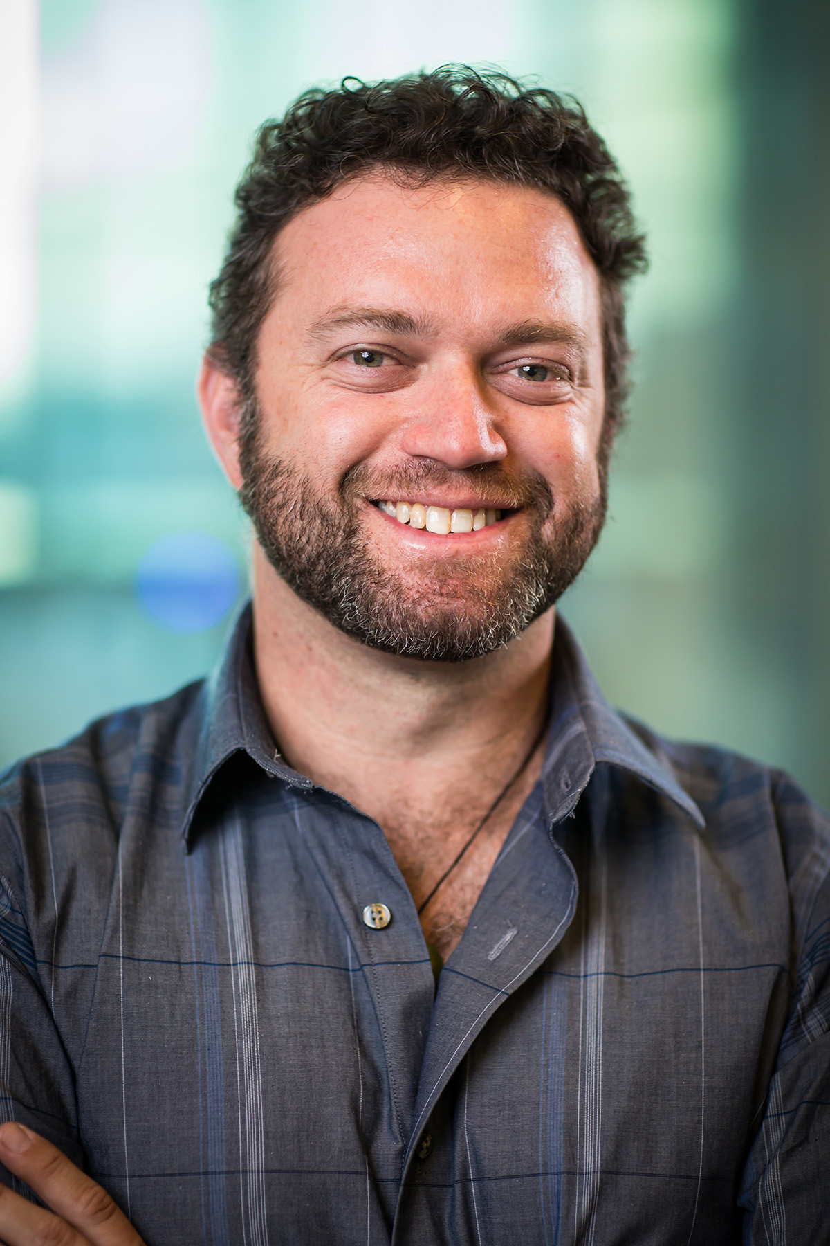
Jason Shepherd, Ph.D., Professor, Spencer Fox Eccles School of Medicine, University of Utah, Salt Lake City, UT
Virus-Like Intercellular Transmission of Tau in Alzheimer’s Disease
Years of research have greatly expanded our understanding of Alzheimer’s Disease, marked by cognitive decline, but much remains to be learned about its causes and how pathology spreads in the brain. Dr. Shepherd and his lab are focused on the role of tau, a protein present in brain cells that can become misfolded and tangled with age. There is a strong correlation between the amount of misfolded tau and cognitive decline in Alzheimer’s disease. To protect cells, misfolded tau needs to be expelled before it builds up to toxic levels and causes cell death. However, misfolded tau released from cells can spread tau pathology to other cells and across the brain.
Precisely how tau is released from cells is unclear, but this may occur as “naked” protein or packaged in membrane wrapped extracellular vesicles (EVs). Shepherd’s team is exploring this second possibility following a new discovery by the lab: that Arc, a neuronal gene critical for synaptic plasticity and memory consolidation, may have evolved from an ancient retrovirus-like element and retained the ability to form EVs by making virus-like capsids that package material and send it to nearby cells. Arc binds Tau, so Arc EVs may also spread the misfolded Tau, contributing to the progression of Alzheimer’s Disease.
In his new research, Shepherd and his team aim to understand the molecular mechanisms of tau release in EVs, the role of Arc in tau pathology, and how Arc-dependent mechanisms contribute to tau spread. Understanding these mechanisms may eventually lead to therapies that reduce the spread of misfolded tau, changing the trajectory of Alzheimer’s Disease pathology.

