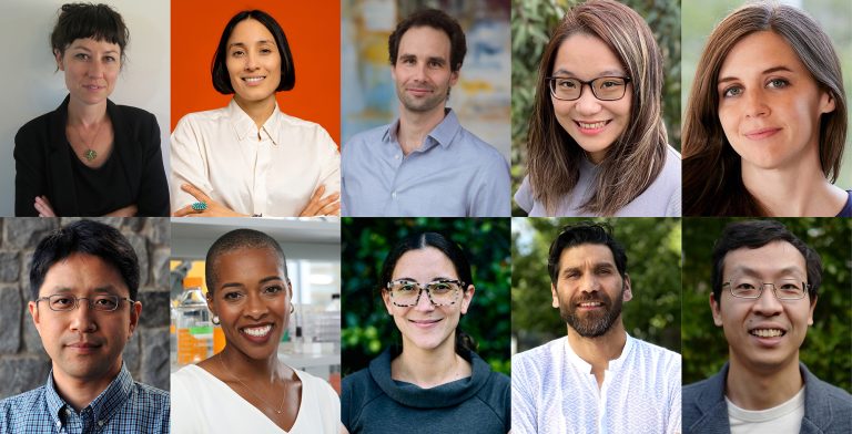
The Board of Directors of The McKnight Endowment Fund for Neuroscience is pleased to announce it has selected six neuroscientists to receive the 2019 McKnight Scholar Award.
The McKnight Scholar Awards are granted to young scientists who are in the early stages of establishing their own independent laboratories and research careers and who have demonstrated a commitment to neuroscience. “The research of this year’s McKnight Scholar awardees exemplifies the spectacular advances that are being made at the cutting edge of neuroscience,” says Kelsey C. Martin M.D, Ph.D, chair of the awards committee and dean of the David Geffen School of Medicine at UCLA. Since the award was introduced in 1977, this prestigious early-career award has funded more than 235 innovative investigators and spurred hundreds of breakthrough discoveries.
“This year’s Scholars tackle brain biology across multiple levels of analysis in a variety of model organisms,” says Martin. “By solving the molecular structure of proteins, elucidating the cell biology of brain cells and dissecting the neural circuits underlying complex behaviors, their discoveries promise to provide insights not only into normal brain function but also into the causes, and potential therapies, of brain disorders. On behalf of the entire committee, I would like to thank all the applicants for this year’s McKnight Scholar Awards for their outstanding scholarship and dedication to neuroscience.”
Each of the following six McKnight Scholar Award recipients will receive $75,000 per year for three years. They are:
| Jayeeta Basu, Ph.D. New York University School of Medicine New York, NY |
Cortical Sensory Modulation of Hippocampal Activity and Spatial Representation – Investigating how different inputs from different brain regions related to space and senses work together to form memories of experiences. |
| Juan Du, Ph.D. Van Andel Research Institute, Grand Rapids, MI |
Regulation mechanism of thermosensitive receptors in nervous system – Researching how different temperature-sensitive receptors in neurons work and how they influence reactions to external heat and cold and internal body temperature. |
| Mark Harnett, Ph.D. Massachusetts Institute of Technology Cambridge, MA |
Perturbing Dendritic Compartmentalization to Evaluate Single Neuron Cortical Computations – Studying how dendrites, the antenna-like input structures of neurons, contribute to computation in neural networks. |
| Weizhe Hong, Ph.D., University of California – Los Angeles Los Angeles, CA |
Neural Circuit Mechanisms of Maternal Behavior – Research into the role of brain circuits in controlling social behaviors, especially the sexually dimorphic functions of these brain circuits and their experience-dependent changes. |
| Rachel Roberts-Galbraith, Ph.D. University of Georgia Athens, GA |
Regeneration of the Central Nervous System in Planarians – A study of central nervous system regeneration in a remarkable species of flatworm, which can regrow its entire nervous system perfectly after nearly any injury. |
| Shigeki Watanabe, Ph.D. Johns Hopkins University Baltimore, MD |
Mechanistic Insights into Membrane Remodeling at Synapses – Investigating the way neurons remodel their membranes within milliseconds for synaptic transmission, critical to the speed with which the nervous system works. |
There were 54 applicants for this year’s McKnight Scholar Awards, representing the best young neuroscience faculty in the country. Young faculty are only eligible for the award during their first four years in a full-time faculty position. In addition to Martin, the Scholar Awards selection committee included Dora Angelaki, Ph.D., New York University; Gordon Fishell, Ph.D., Harvard University; Loren Frank, Ph.D., University of California, San Francisco; Mark Goldman, Ph.D., University of California, Davis; Richard Mooney, Ph.D., Duke University School of Medicine; Amita Sehgal, Ph.D., University of Pennsylvania Medical School; and Michael Shadlen, M.D., Ph.D., Columbia University.
Applications for next year’s awards will be available in September and are due in early January 2020. For more information about McKnight’s neuroscience awards programs, please visit the Endowment Fund’s website at https://www.mcknight.org/programs/the-mcknight-endowment-fund-for-neuroscience
About The McKnight Endowment Fund for Neuroscience
The McKnight Endowment Fund for Neuroscience is an independent organization funded solely by the McKnight Foundation of Minneapolis, Minnesota and led by a board of prominent neuroscientists from around the country. The McKnight Foundation has supported neuroscience research since 1977. The Foundation established the Endowment Fund in 1986 to carry out one of the intentions of founder William L. McKnight (1887-1979). One of the early leaders of the 3M Company, he had a personal interest in memory and brain diseases and wanted part of his legacy used to help find cures. The Endowment Fund makes three types of awards each year. In addition to the McKnight Scholar Awards, they are the McKnight Technological Innovations in Neuroscience Awards, providing seed money to develop technical inventions to enhance brain research; and the McKnight Memory and Cognitive Disorders Awards, for scientists working to apply the knowledge achieved through basic research to human brain disorders that affect memory or cognition.
2019 McKnight Scholar Awards
Jayeeta Basu, Ph.D., Assistant Professor, Neuroscience Institute,
New York University School of Medicine, New York, NY
Cortical Sensory Modulation of Hippocampal Activity and Spatial Representation
The brain can store a lot of information in a memory, including where it happened and under what sensory context such as sights, sounds, smells, rewards or punishments. Exactly how those different pieces of information are linked to form episodic memories, and how these memories can be recalled instantaneously from cues in the future is the basis of Dr. Basu’s research. In particular, Dr. Basu and her team will investigate the relationship between the entorhinal cortex and the hippocampus in forming memories about places.
Two parts of the entorhinal cortex deliver different inputs. The medial entorhinal cortex (MEC) shares spatial information like direction, distance and orientation, while the lateral entorhinal cortex (LEC) delivers contextual information from the senses, including smell, sound, novelty, and objects. Inputs from both are delivered to the hippocampus and help to form crucial memories of places stored in specific groups of “place cells” in the brain, such as where to find food, or areas to avoid because predators are present. Critically, these memories of place and the cognitive map of space need to be on the one hand stable in the face of environmental changes such as weather or time of day yet on the other hand flexible, since food or predators may move. Little is understood of what information is enough and necessary to create, maintain and change these memories, especially how these are shaped by sensory information from the LEC in association with spatial information from MEC.
Dr. Basu aims to map the circuitry involved between the LEC and specific hippocampal neurons. Her lab will directly record the signals received by the thin dendrites of the neurons when LEC signals are sent with or without MEC signals, and at different signal strengths. A second series of experiments with mice will test the hypothesis that these LEC inputs support the creation of memories of place while learning – scent cues will trigger behavior to seek rewards at distinct places. Researchers will see how switching on or off LEC signals during learning or during recall affect the activation of place cells in the brain and the learning behavior itself. This research may be relevant in future studies of Alzheimer’s disease, PTSD and other conditions where memory and contextual “triggers” are activated.
Juan Du, Ph.D., Assistant Professor, Structural Biology Program, Center for Cancer and Cell Biology, Van Andel Research Institute, Grand Rapids, MI
- https://dulab.vai.org/
Regulation mechanism of thermosensitive receptors in nervous system
When it comes to sensing and reacting to changes in temperature, both external and internal, little is known of the exact mechanism and process. Ion channel receptors on neurons open or close to allow signals to pass through, and these channels can be activated by chemicals, mechanical processes or temperature, but what exactly about temperature that makes temperature-activated channels activate is unclear.
Dr. Du will conduct a three-part project to unlock the secrets of how temperature information is received and processed by the neural system. She is looking at three particular receptors, one that detects cool and cold temperatures externally, one that detects extreme external heat, and one that detects warm temperatures in the brain (for regulating body temperature.) She will first identify purification conditions for these receptors so they can be extracted and used in lab experiments and still operate the same as receptors in the body.
A second aim is to see what structures on the receptors are activated by temperature and understanding how they work. This will also include the development of new therapeutics that can bind to these structures and regulate them. Third, when the structures are understood, validation experiments in which the receptors are mutated to change or remove temperature sensitivity will be conducted, first on cells, and then in mice, to see how alterations to temperature-sensitive receptors impact behavior. Once the function and regulation of these receptors is understood, it can open the way to treatments for certain neurodegenerative diseases, temperature-related conditions and even pain management, since some temperature-sensitive sensors are related to pain transmission.
Mark Harnett, Ph.D., Assistant Professor, Brain and Cognitive Sciences, Massachusetts Institute of Technology, Cambridge, MA
Perturbing Dendritic Compartmentalization to Evaluate Single Neuron Cortical Computations
The brain can process and act on an amazing amount of information because of the way neurons are networked together. There is still more to be learned, however, about how neurons themselves work. Dr. Harnett is researching the role dendrites – the tree-like structures that extend from neurons where signals from other neurons are received – to determine whether these substructures themselves give individual neurons the power to perform more complex computations than generally believed.
Conventional wisdom is that neurons take in data from other neurons, and if the data reach a certain threshold, the neuron fires, passing on the information. Dr. Harnett is investigating how the dendrites themselves might also be filtering or boosting signals. Some branches are closer to the soma (the output part of the neuron) than others, so which branch receives a signal may impact the effect of the signal. Also, some branches of dendrites seem to be wired to look for and amplify particular types of signals – for example, one branch might specialize in passing on signals for fast-moving, high contrast visual stimuli, but not other stimuli.
Dr. Harnett is looking at dendrites in the visual system with precise electrical and optical tools, to measure how signals travel down dendrite branches, and measure how altering the dendrites changes how the neuron operates. These perturbations will allow Dr. Harnett to test if inhibiting signals on a specific branch of a dendrite changes how the neural network responds to certain visual stimuli. Learning that a single neuron is essentially made up of its own network of smaller signal processors would change our understanding of how the brain computes. Among other things, this could affect how artificial intelligence, which is modelled on neural networks, evolves in years to come.
Weizhe Hong, Ph.D., Assistant Professor, Departments of Biological Chemistry and Neurobiology, University of California, Los Angeles, CA
Neural Circuit Mechanisms of Maternal Behavior
Many social behaviors exhibit striking sex differences in their levels and forms and undergo experience-dependent changes throughout the animals’ lives. One prominent example is parenting behavior, which is a prevalent social behavior shared across the animal kingdom from invertebrates to humans and is critical for offspring survival. Parenting behavior often differs greatly between males and females and can undergo drastic changes as animals mature and give birth. However, the brain circuits underlying parenting behavior and its differences between sexes and physiological states are not well defined.
A particular focus of Dr. Hong’s work will be investigating the role of an evolutionarily conserved brain region called the amygdala in controlling parenting behavior. While female mice usually engage in extensive pup nurturing behaviors, male mice generally do not show parenting behavior until their own offspring are born. The sex differences and physiological switch in mouse parenting behavior provide an excellent opportunity to understand the neural mechanisms underlying sexually dimorphic display of parenting behavior and its physiological-state-dependent transitions.
The research will identify specific, molecularly defined neuronal populations that mediate parenting behavior. The research will also compare the neural circuits in males and females to understand how neural activity in these neurons regulates parenting behavior. This research will provide key insights into the neural basis of an essential social behavior and the basic principles governing sexually dimorphic behaviors. Such insights may also improve our understanding of the regulation of human parental and social behaviors in both health and disease.
Rachel Roberts-Galbraith, Ph.D., Assistant Professor, Department of Cellular Biology, University of Georgia, Athens, GA
Regeneration of the Central Nervous System in Planarians
Developing a central nervous system in an animal is a remarkably complex process. Regenerating a damaged neural system is even more complicated, since it requires activating all the same development processes in one area but not another and rewiring neurons so they will work as they did before. Humans have very poor central nervous system regenerative ability, so damage to the brain or spinal cord is often irreversible. Dr. Roberts-Galbraith hopes to understand more about how neural repair can work by researching regeneration in planarians, a remarkable type of flatworm that can regrow its entire central nervous system (and the rest of its body) even after dramatic injuries.
By studying successful neural regeneration in the natural world, Dr. Roberts-Galbraith hopes to learn details about the mechanism of neural regeneration and the role of different cells. One aim is to investigate whether neurons can detect injury and self-initiate repairs themselves by sending signals that trigger and direct regrowth. Dr. Roberts-Galbraith hypothesizes that neurons influence planarian stem cells, which are recruited to regrow parts of the central nervous system (and other body parts). Fine control of stem cells is critical for regeneration, as planarians faithfully replace missing tissues and never develop tumors.
Another aim is to examine the role of glial cells, which have traditionally been seen as the glue of the nervous system but which clearly possess more significant roles than previously recognized. Glial cells make up a large part of animals’ nervous systems and must be regenerated along with neurons; they are also likely to modulate neuronal regeneration. The hope is this research will provide more understanding of how regeneration can happen in the most successful cases, and perhaps inform new ways of thinking about neural regeneration in humans.
Shigeki Watanabe, Ph.D., Assistant Professor of Cell Biology and Neuroscience, Johns Hopkins University, Baltimore, MD
Mechanistic Insights into Membrane Remodeling at Synapses
The lightning-fast speed of neural networks allows us to sense, evaluate and react to the world around us. It also has required neurons to develop some remarkable properties. In his research, Dr. Watanabe will investigate one of the most remarkable – the ability of neurons to remodel their membranes on a millisecond timescale for neuronal communication using processes not fully understood.
The membrane around a neuron needs to adapt to allow the neuron to grow, migrate and – importantly – allow other membranes to merge and separate during neuronal communication. In the process under investigation, a “bubble” of membrane called the synaptic vesicle merges with the neuronal membrane, after which a new piece of membrane essentially bulges inward and pinches off. The mechanism believed to be used, clathrin-mediated endocytosis, just isn’t fast enough to allow these vesicles to be created and recycled on the timescale in which synaptic transmission happens. Dr. Watanabe discovered a new mechanism, ultrafast endocytosis, that handles the process, but understanding just how it works has been hampered by the small size of synapses and rapid speed of this process.
Dr. Watanabe will use a technique called flash-and-freeze electron microscopy to research this process. Neurons will be stimulated with light – the flash – then the process will be stopped precisely with high-pressure freezing at precise time intervals microseconds after stimulation. The frozen synapses can then be visualized with an electron microscope. By taking a series of images frozen at different time intervals after stimulation, Dr. Watanabe will create a step-by-step visualization of the process and identify the proteins involved and what they do. Not only will this give a better understanding of how neurons work, it has implications for diseases that are related to faulty neural transmission, such as Alzheimer’s Disease.


