December 3, 2019
The McKnight Endowment Fund for Neuroscience has selected four projects to receive the 2020 Memory and Cognitive Disorders Awards. The awards will total $1.2 million over three years for research on the biology of brain diseases, with each project receiving $300,000 between 2020 and 2023.
The Memory and Cognitive Disorders (MCD) Awards support innovative research by U.S. scientists who are studying neurological and psychiatric diseases, especially those related to memory and cognition. The awards encourage collaboration between basic and clinical neuroscience to translate laboratory discoveries about the brain and nervous system into diagnoses and therapies to improve human health.
“We are thrilled to select some of the best scientists and their work in the country this year,” said Ming Guo, M.D., Ph.D., chair of the awards committee and Professor in Neurology & Pharmacology at UCLA David Geffen School of Medicine. “These scientists are addressing questions related to how general anesthesia and sleep impact memory, and how memory works at the basic level. Together, we aim to understand the underlying neurobiology of memory and brain disorders that one day will translate into cures of some of the most devastating brain disorders that afflict millions of people in the world.”
The awards are inspired by the interests of William L. McKnight, who founded The McKnight Foundation in 1953 and wanted to support research on diseases affecting memory. His daughter, Virginia McKnight Binger, and The McKnight Foundation board established the McKnight neuroscience program in his honor in 1977.
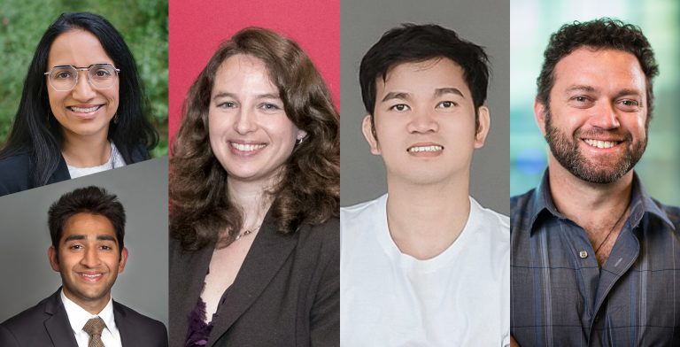
Up to four awards are given each year. This year’s awardees are:
Ehud Isacoff, Ph.D., Evan Rauch Chair, Department of Neuroscience, University of California, Berkeley; and Dirk Trauner, Ph.D. Janice Cutler Chair in Chemistry and Adjunct Professor of Neuroscience and Physiology, New York University – Photo-activation of Dopamine Receptors in Models of Parkinson’s Disease: Dr. Isacoff and Dr. Trauner are investigating whether specialized-designed photo-sensitive molecules can be introduced into the brains of a mice (whose dopamine reception has been impaired in a way that resembles Parkinson’s Disease) and have their cognitive function restored through light activation.
Mazen Kheirbek, Ph.D., Assistant Professor of Psychiatry, Center for Integrative Neuroscience, University of California, San Francisco; and Jonah Chan, Ph.D., Professor of Neurology, Weill Institute for Neurosciences, University of California, San Francisco – New Myelin Formation in Systems Consolidation and Retrieval of Remote Memories: The research of Dr. Kheirbek and Dr. Chan explores why some memories are easier to recall than others; the focus is on the differing development of myelin sheaths around the axons of some neurons during contextual conditioning.
Thanos Siapas, Ph.D., Professor of Computation and Neural Systems, Division of Biology and Biological Engineering, California Institute of Technology – Circuit Dynamics and Cognitive Consequences of General Anesthesia: Dr. Siapas seeks to gain a deeper understanding of how general anesthesia works and how it affects the brain; for the project he plans to record brain activity from anaesthetized mice and use machine learning to uncover patterns, as well as study the long-term effects of anesthesia.
Carmen Westerberg, Ph.D., Associate Professor, Department of Psychology, Texas State University; and Ken Paller, Ph.D. Professor of Psychology and James Padilla Chair in Arts & Sciences, Department of Psychology, Northwestern University – Does Superior Sleep Physiology Contribute to Superior Memory Function? Implications for Counteracting Forgetting: Dr. Westerberg and Dr. Paller are exploring the role of sleep in memory consolidation by studying individuals with highly superior autobiographical memory. Analyzing how their sleep differs from the general population may enable future research benefitting those who are suffering memory loss.
With 100 letters of intent received this year, the awards are highly competitive. A committee of distinguished scientists reviews the letters and invites a select few researchers to submit full proposals. In addition to Dr. Guo, the committee includes Sue Ackerman, Ph.D., University of California, San Diego; Susanne Ahmari, M.D., Ph.D., University of Pittsburgh School of Medicine; Robert Edwards, M.D., University of California, San Francisco; Harry Orr, Ph.D., University of Minnesota; Steven E. Petersen, Ph.D., Washington University in St. Louis; and Matthew Shapiro, Ph.D., Albany Medical Center.
Letters of intent for the 2021 awards are due by March 2, 2020.
About the McKnight Endowment Fund for Neuroscience
The McKnight Endowment Fund for Neuroscience is an independent organization funded solely by The McKnight Foundation of Minneapolis, Minnesota, and led by a board of prominent neuroscientists from around the country. The McKnight Foundation has supported neuroscience research since 1977. The Foundation established the Endowment Fund in 1986 to carry out one of the intentions of founder William L. McKnight (1887–1978), one of the early leaders of the 3M Company.
The Endowment Fund makes three types of awards each year. In addition to the Memory and Cognitive Disorders Awards, they are the McKnight Technological Innovations in Neuroscience Awards, providing seed money to develop technical inventions to advance brain research; and the McKnight Scholar Awards, supporting neuroscientists in the early stages of their research careers.
2020 McKnight Memory and Cognitive Disorders Awards
Ehud Isacoff, Ph.D., Evan Rauch Chair, Department of Neuroscience, University of California, Berkeley; and Dirk Trauner, Ph.D. Janice Cutler Chair in Chemistry and Adjunct Professor of Neuroscience and Physiology, New York University
“Photo-activation of Dopamine Receptors in Models of Parkinson’s Disease”
Dopamine is generally known for its association with creating positive sensations or for its role in addiction. But in fact, dopamine plays a wide range of roles, and there are five different types of dopamine receptors found in brain cells, each of which has many complicated downstream effects relating to movement, learning, sleep and more. In addition to being a movement disorder, Parkinson’s disease is also a cognitive disorder and is brought on by a loss of dopamine input.
Drs. Isacoff and Trauner are exploring new ways to precisely control dopamine receptor activation in brains that mimic the loss of reception found in Parkinson’s patients. The lab’s approach uses a synthetic photoswitchable tethered ligand (PTL) – essentially, a dopamine mimic attached by a leash to an anchor, which in turn will attach only to specific dopamine receptors in specific cells. The PTLs are introduced into the brain, and optical wires deliver light pulses directly to the areas where the PTLs are, similar to the setup used to deliver electrical impulses in deep brain stimulation. The experiments will observe if animals that have had dopamine signaling knocked out can regain movement control using targeted PTLs and light – instantly, precisely reactivating function with the flip of a switch, without the unintended side effects of pharmacological fixes.
The research conducted by Drs. Isacoff and Trauner will perfect the process of developing and delivering these PTLs and potentially demonstrate their effectiveness. This could result in a new class of treatments not only for Parkinson’s, but potentially other brain disorders as well.
Mazen Kheirbek, Ph.D., Assistant Professor of Psychiatry, Center for Integrative Neuroscience, University of California, San Francisco; and Jonah Chan, Ph.D., Professor of Neurology, Weill Institute for Neurosciences, University of California, San Francisco
“New Myelin Formation in Systems Consolidation and Retrieval of Remote Memories”
The brain physically changes as it takes in and stores data – as if you opened a computer after saving data and found that a wire had grown thicker or extended to a nearby circuit as well. This process notably occurs in the formation of myelin sheathes around axons (a part of neurons) which has been shown to play a role in increased efficiency of communication within and between neuronal circuits, which may facilitate the recall of some memories.
What isn’t understood is whether these sheathes form around axons related to some memories more than others. Using a mouse model, Dr. Kheirbek and Dr. Chan are exploring this process, seeking to understand if the axons of neuronal ensembles activated by fearful experiences are preferentially myelinated – essentially, making traumatic memories easier to recall – and how this process works and can be manipulated. Preliminary research found that fear conditioning resulted in an increase in cells that are precursors for myelin formation, and that this process was involved in the long-term consolidation of fear memories.
One experiment will tag which cells are activated during contextual fear conditioning and observe myelination in those cells; then, the researchers will manipulate the electrical activity of distinct circuits to determine what causes the additional myelination to occur. Additional experiments will observe if mice that have had new myelin formation suppressed exhibit the same fear responses as mice with normal myelin formation. A third experiment will observe the entire process with high-resolution live imaging over a long period. The research could have implications for conditions such as Post Traumatic Stress Disorder, where traumatic memories and fear response are activated, or memory disorders where recall is disturbed.
Thanos Siapas, Ph.D., Professor of Computation and Neural Systems, Division of Biology and Biological Engineering, California Institute of Technology
“Circuit Dynamics and Cognitive Consequences of General Anesthesia”
While general anesthesia (GA) has been a boon to medicine by allowing surgeries that would be impossible in awake patients, the exact ways GA affects the brain and its long-term effects are poorly understood. Dr. Siapas and his team are looking to expand our fundamental knowledge of GA effects on the brain in a series of experiments, opening the door for additional research into the function and application of GA that could someday lead to its improved use in humans.
Dr. Siapas aims to use multielectrode recordings to monitor brain activity during anesthesia, and to employ machine learning approaches to detect and characterize patterns in the neural data. The team will record activity during induction and emergence from GA, as well as during steady state, to determine exactly what states the brain goes through. This research may be especially useful in understanding and helping prevent interoperative awareness, a situation where patients sometimes become aware of what is happening but unable to move, which can lead to severe trauma.
A final experiment will look at the long-term cognitive impact of GA. Many people experience short-term cognitive impacts after anesthesia, but a small percentage suffer long-term or permanent cognitive impairment. The team will manipulate GA administration (again in mice), then test for deficits in learning or cognition, and record the brain activity associated with these deficits.
Carmen Westerberg, Ph.D., Associate Professor, Department of Psychology, Texas State University; and Ken Paller, Ph.D., Professor of Psychology and James Padilla Chair in Arts & Sciences, Department of Psychology, Northwestern University
“Does Superior Sleep Physiology Contribute to Superior Memory Function? Implications for Counteracting Forgetting”
Drs. Westerberg and Paller and their team hope to gain insight into the process of forgetting by studying the sleep physiology of people who almost never forget. These individuals, who have a condition called “highly superior autobiographical memory,” or HSAM, can effortlessly remember the minute details of every day of their lives with equal clarity, whether it happened last week or 20 years ago. By comparison, most humans can remember the same amount of detail as those with HSAM for some weeks, but beyond that they recall only highly significant moments in detail.
Sleep physiology is proposed as one possible difference between those with HSAM and those without. Sleep is known to play an important role in memory consolidation, and a detailed human study of the brain activity during sleep of HSAM and control individuals will record, compare and analyze the patterns of slow oscillations (linked to memory consolidation), sleep spindles (also connected to consolidation, and recorded at high levels in HSAM individuals) and the ways in which they co-occur.
A second study features an easy-to-use headband that will allow subjects to measure both sleep and memory data at home over a one-month period, to determine if enhanced sleep physiology over multiple nights contributes to superior memory for events that happened one month prior. In addition, by guiding the reactivation of memories that are not autobiographical in nature with sound cues presented during sleep, this study will help reveal whether enhanced sleep physiology in HSAM individuals can enhance memory for non-autobiographical memories as well. Drs. Westerberg and Paller hope that by finding how highly superior memory works, we might be able to uncover patterns in those suffering from sub-optimal memory function, such as those suffering from Alzheimer’s Disease, and perhaps find new ways to understand and treat the conditions.


